

Home > Company News
Nasal bleeding may arise from an easily visible area at the front of the septum, but it can also originate from the posterior nasal cavity, the deeper inferior meatus, or the olfactory cleft. Blood, clots, and narrow anatomy can hide the true source during a limited anterior examination. A nasal endoscope provides illumination, magnification, and angled views, allowing the clinician to clear the field, inspect the nasal cavity systematically, and confine treatment as closely as possible to the bleeding site.
This locate-first, treat-second approach can reduce the pain and mucosal injury associated with broad blind packing. It is particularly helpful in recurrent bleeding, suspected posterior epistaxis, or bleeding that returns after previous packing.
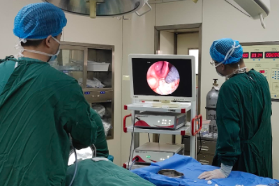
• Assess blood loss, vital signs, medications, and relevant medical conditions; start resuscitation, laboratory testing, or emergency care when indicated.
• Under monitoring and adequate topical anesthesia, remove clots and improve visualization. A clinician may use a topical vasoconstrictor when appropriate.
• Inspect the septum, floor, inferior and middle meatuses, posterior choana, and nasal roof in an orderly fashion for active bleeding, a vascular stump, or another lesion.
• Select the most focused treatment based on vessel type, location, mucosal condition, and the patient's overall risk.
Targeted micro-packing is useful when the source is known but immediate cautery is unsuitable. Under direct endoscopic vision, an absorbable hemostatic agent or a small packing material can be placed precisely near the bleeding point, limiting the effect on the rest of the nasal airway.
Chemical cautery or bipolar electrocautery may be used for a clearly defined superficial vessel or small vascular stump. The aim is not to create a larger treatment field. The clinician confirms the source, applies limited coagulation, and avoids deep injury to corresponding areas on both sides of the septum.
Radiofrequency, coblation, laser, or other energy devices may be selected for specific indications and equipment. Output and application time should follow the device instructions, tissue response, and clinical judgment rather than a universal fixed setting. Persistent high-volume posterior bleeding may require sphenopalatine artery control, another surgical ligation procedure, or endovascular embolization.
• Energy-based hemostasis must be performed by trained clinicians with attention to thermal injury, crusting, adhesions, and septal perforation.
• Patients taking anticoagulants or antiplatelet drugs should not stop them on their own; the prescribing clinician and ENT team should coordinate the decision.
• Hypertension, blood disorders, liver or kidney disease, and recent nasal surgery may increase complexity and require individualized monitoring.
• Recurrent unilateral bleeding, nasal obstruction, or an abnormal bulge warrants evaluation for a vascular lesion, mass, or another local cause.
Urgent assessment is appropriate when heavy bleeding continues despite firm compression, blood keeps running into the throat, dizziness, weakness, or breathing difficulty develops, bleeding follows significant trauma, or bleeding is difficult to control in a person taking anticoagulants. Endoscopic hemostasis is a medical procedure and should never be attempted at home.

+86-13167065777
Building 5A, No. 9628, Hunan Highway, Pudong New Area, Shanghai, China
 Links:
Shanghai Shiyin Photoelectric Instrument Co.,Ltd. All Right Reserved
